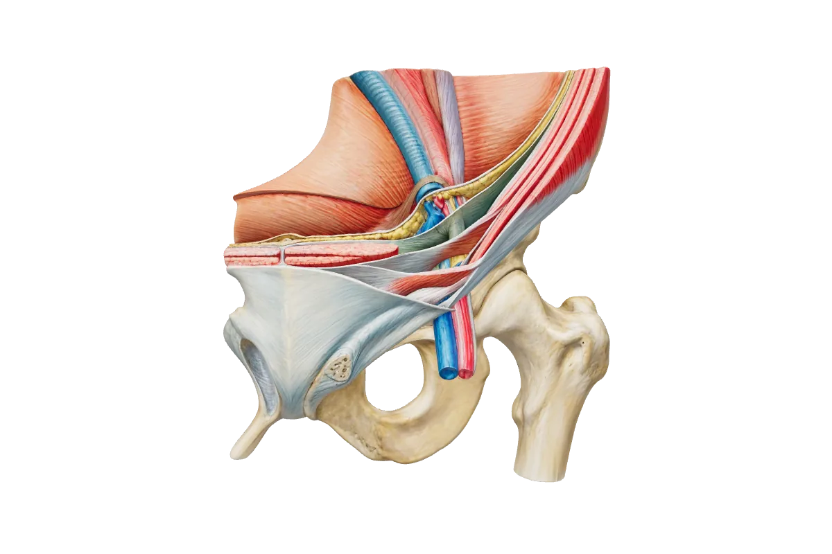
The inguinal canal and groin hernia anatomy, open and laparoscopic
Groin anatomy for FRCS (Gen Surg): the walls and rings of the inguinal canal, direct, indirect and femoral hernias, the nerves at risk, and the laparoscopic triangles of doom and pain.
The inguinal canal is an oblique passage about 4 cm long above the medial half of the inguinal ligament. It carries the spermatic cord in men and the round ligament of the uterus in women, and the ilioinguinal nerve in both. Its obliquity, the shutter action of the internal oblique and transversus, and the strength of the posterior wall medially protect it against herniation.
The boundaries of the inguinal canal.
Wall
Formed by
Anterior
The external oblique aponeurosis, reinforced laterally by the internal oblique
Posterior
The transversalis fascia, reinforced medially by the conjoint tendon
Roof
The arching fibres of the internal oblique and transversus abdominis
Floor
The inguinal ligament, with the lacunar ligament medially
The deep (internal) ring is an opening in the transversalis fascia at the midpoint of the inguinal ligament, about 1.5 cm above the femoral pulse (at the mid-inguinal point), immediately lateral to the inferior epigastric vessels.
The superficial (external) ring is a triangular defect in the external oblique aponeurosis, above and medial to the pubic tubercle.
Interactive 3D · Inguinal canal & hernia anatomy. The left groin from the front: find the deep ring, then the inguinal ligament with the femoral artery, vein and canal beneath it.Open full screen
The spermatic cord
The cord carries the vas deferens, the testicular artery, the artery to the vas and the cremasteric artery, the pampiniform plexus, the genital branch of the genitofemoral nerve, sympathetic fibres, lymphatics and the remnant of the processus vaginalis. It gathers three coverings as it passes through the abdominal wall: the internal spermatic fascia from the transversalis fascia, the cremasteric fascia and muscle from the internal oblique, and the external spermatic fascia from the external oblique. The ilioinguinal nerve runs on the cord, outside these coverings, and leaves through the superficial ring; it does not pass through the deep ring.
Direct, indirect and femoral hernias
Only at operation (or on imaging) can direct and indirect hernias be reliably told apart.
Row
Indirect inguinal
Direct inguinal
Femoral
Route
Through the deep ring, within the cord
Through the posterior wall in Hesselbach’s triangle
Through the femoral canal, beneath the inguinal ligament
Relation to the inferior epigastric vessels
Lateral
Medial
Below and lateral to the pubic tubercle
Origin
A patent processus vaginalis
Weakness of the transversalis fascia
Widening of the femoral ring
Typical patient
Any age; the usual hernia of children
Older men
Older women, though inguinal hernias are still commoner in women
Hesselbach’s triangle is bounded by the inferior epigastric vessels laterally, the lateral border of rectus abdominis medially and the inguinal ligament below. A pantaloon hernia has both components, straddling the vessels.
The femoral canal is the medial compartment of the femoral sheath. Its ring is bounded by the inguinal ligament in front, the pectineal (Cooper’s) ligament behind, the lacunar ligament medially and the femoral vein laterally. The rigid medial and anterior borders explain why femoral hernias strangulate, why they should be repaired promptly rather than watched, and why the lacunar ligament may have to be divided to reduce one — taking care of an abnormal obturator artery that can run across it.
Nerves at risk in open repair
Ilioinguinal nerve (L1): on the cord beneath the external oblique aponeurosis; supplies the skin of the upper medial thigh and the root of the penis or labium majus.
Iliohypogastric nerve (L1): between the internal and external oblique above the canal; supplies the suprapubic skin.
Genital branch of the genitofemoral nerve (L1–L2): within the cord, with the cremasteric vessels; supplies cremaster and scrotal skin.
Identifying and preserving these nerves, and avoiding entrapment by mesh fixation, is the main way to prevent chronic postoperative inguinal pain. The HerniaSurge guidelines recommend identifying all three in open repair, and pragmatic neurectomy of a nerve already at risk rather than routine division.
The laparoscopic view
From inside, the landmarks are the inferior epigastric vessels, the medial umbilical ligament, the vas deferens running medially and the gonadal vessels laterally, converging on the deep ring. The iliopubic tract is the posterior counterpart of the inguinal ligament.
Triangle of doom
Between the vas deferens medially and the gonadal vessels laterally, with its apex at the deep ring. It contains the external iliac artery and vein: no tacks or dissection here.
Triangle of pain
Lateral to the gonadal vessels and below the iliopubic tract. It contains the lateral femoral cutaneous nerve, the femoral branch of the genitofemoral nerve and, deeper, the femoral nerve: no tacks below the iliopubic tract laterally.
Corona mortis
An anastomosis between the obturator and inferior epigastric or external iliac vessels over the superior pubic ramus, at risk when dissecting onto Cooper’s ligament.
Interactive 3D · Femoral triangle & canal. The femoral triangle and canal (in preparation).Open full screen
A fit 52-year-old woman has a symptomatic, reducible lump in the right groin. On examination it appears to be an inguinal hernia, and ultrasound reports a fat-containing groin hernia without specifying the type.
Which is the most appropriate repair?
How sure are you?
Explanation
The principle is that a groin hernia in a woman should be repaired by a method that covers the whole myopectineal orifice. Femoral hernias are much more frequent in women, are often mistaken for inguinal hernias before operation, and account for a large share of 'recurrences' after open anterior repair in women, many of which are femoral hernias that were missed. A laparoscopic preperitoneal mesh (TEP or TAPP) inspects and covers the inguinal and femoral orifices together and is recommended by the international guidelines for all women. Lichtenstein, Shouldice and plug repairs leave the femoral canal unprotected. Watchful waiting is not advised in women because of the higher risk of a femoral hernia and of strangulation. Reference: HerniaSurge Group, International guidelines for groin hernia management, Hernia 2018.
How do you tell a direct from an indirect inguinal hernia?+
By their relation to the inferior epigastric vessels: indirect hernias pass through the deep ring lateral to them, direct hernias through Hesselbach’s triangle medial to them. Clinical tests are unreliable; the distinction is made at operation or on imaging.
What are the boundaries of the femoral canal?+
The inguinal ligament in front, the pectineal ligament behind, the lacunar ligament medially and the femoral vein laterally.
Which nerve is injured if a patient has numbness over the upper medial thigh after open hernia repair?+
The ilioinguinal nerve, which runs on the spermatic cord in the canal and supplies the skin of the upper medial thigh and the root of the penis or labium majus.
What is the triangle of doom?+
In laparoscopic repair, the triangle between the vas deferens and the gonadal vessels, with its apex at the deep ring, containing the external iliac vessels.
References
HerniaSurge Group. International guidelines for groin hernia management. Hernia 2018;22(1):1–165.
Standring S (ed). Gray’s Anatomy: The Anatomical Basis of Clinical Practice. 42nd ed. Elsevier; 2020.
O’Connell PR, McCaskie AW, Sayers RD (eds). Bailey & Love’s Short Practice of Surgery. 28th ed. CRC Press; 2023.
Written for revision in the FRCS (Gen Surg) style under our editorial standards. It is not clinical advice: always confirm against current guidance. Spotted an error? Tell us.